Each hip bone consists of an ilium ischium and pubis. Label the following structures on the lateral view of the pelvis below.

Coxal Pelvic Bone Posterior View With Labels Appendicular Skeleton Visual Atlas Page 18 Anatomy Flashcards Medical Anatomy Anatomy And Physiology
Bones of the pelvis by mdanielsmelear14 354 plays 9p Image Quiz.

. After you have studied the bones in lab label the drawings as a self-test. Sacroiliac joint articulation with the sacrum. The hip bones have three main articulations.
You need to be a group member to play the tournament. The hip bone has three parts. The left and right hip bones innominate bones pelvic bones are two irregularly shaped bones that form part of the pelvic girdle the bony structure that attaches the axial skeleton to the lower limbs.
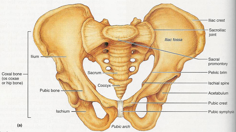
These bony components are the ilium ischium and pubis. Os means bone and coxae means of the hip so its the bone of the hip the hip bone. Superior view of female pelvis and surrounding endopelvic fascia.
2 coxal or coxa hip bones unite with the sacrum. Posterior inferior iliac spine anterior superior iliac spine superior pubic ramus anterior gluteal line Solutionpdf Experts Answer. Use the images above to answer questions 1-25.
Each adult hip bone is formed by three separate bones that fuse together during the late teenage years. Its also called the greater pelvis. Identify the structure labeled as 1.
Learn vocabulary terms and more with flashcards games and other study tools. There are three bones of the pelvis. The femur attaches to the acetabulum so that structure faces inferior lateral.
Label the following bones. The labels on the left-hand side of the labelled diagram of the left knee order-top to bottom. The figure below is a lateral view of the head.
Ischium ilium ischial tuberosity greater sciatic notch posterior inferior iliac spine iliac crest posterior superior iliac spine ischial spine lesser sciatic notch obturator foramen acetabulum pubic tubercle ilium symphysis pubis public arch acetabulum sacrum sacral promontory ischium sacroiliac joint. Lable the pelvis by USSUMW_Hayley 484 plays 11p Image Quiz. Weve got two hip bones a sacrum and a coccyx.
Do not spend your. After you have labeled the bones coloring them using the following chart. The sacrum and two innominate bones.
You may also find sacrospinous ligament lesser sciatic foramen sacrotuberous ligament ischial tuberosity deep posterior. Bony pelvis or pelvic skeleton is formed by hip bones sacrum and coccyx. Coxal Pelvic bone anterior view with labels - Appendicular Skeleton Visual Atlas page 17 This is Page 17 of a photographic atlas I created as a laboratory study resource for my BIOL 121 Anatomy and Physiology I students on the bones and bony landmarks of.
Learn vocabulary terms and more with flashcards games and other study tools. Canine pelvis x-ray 3 by GracynVH 388 plays 15p Image Quiz. Start studying Lab 17.
Anatomy and Physiology questions and answers. Posterior Superior Ilium Spine. How to determine left and right coxa.
The Living Fabric 5 The Integumentary System 6 Bones And Skeletal Tissues 7 The Skeleton 8 Joints 9 Muscles And Muscle Tissue 10 The Muscular System 11. These bones connect the axial skeleton to the lower limbs and therefore play a role in bearing the weight of the upper body. Formed by the left and right hip bones the pelvic girdle connects the lower limb leg bones to the axial skeleton.
Squamous portion 6 Parietal bone Petrous portion Ethmoid bone Frontal bone Sphenoid bone Temporal bone Occipital bone Correctly label the bones and anatomical features of the pelvic girdle. Each pelvic bone hip bone is made by the combination three bones namely the ilium pubis and ischium. Start studying pelvic girdle label.
The ilium pubis. Study from the bone list or your textbook after you marked the drawings as instructed on page 6-2. The femur is the largest bone in the body and the only bone of the thigh femoral region.
The Living Units 4 Tissue. Structures shown on the lateral side of the uterus from anterior to posterior. The pelvis is a ring structure made up of three bones.
The pelvis consists of two hip bones attached at the front anterior by the pubic symphysis and at the back posterior by the sacrum. Mandible light green temporal bone dark blue. Pelvis by DHS Science 372 plays 10p Image Quiz.
BONES OF THE AXIAL AND APPENDICULAR SKELETON. This game is part of a tournament. Pubic symphysis articulation between the left and right hip bones.
Learn the bones of the pelvic bone. Sacrum is part of vertebrae coxa single bone. Round ligament of the uterus uterine tube ovarian ligament ureter uterosacral ligament.
The answer is B. The hip bone or coxal bone forms the pelvic girdle portion of the pelvis. These bones also act as attachments for many muscles and ligaments within the pelvis and lower limbs.
The paired hip bones are the large curved bones that form the lateral and anterior aspects of the pelvis. The three bones and three joints composing the pelvic ring have no inherent stability without vital ligamentous. These three bones fuse at a cup-shaped concavity called the acetabulum which articulates with the head of the femur to form the hip joint.
The hip bone sacrum and coccyx. Identify the bones and their landmarks on this posterior view of the pelvic girdle. Most but not all features you are required to know are shown on the following pages.
These names are retained and used to. 1 The Human Body. Femur- This is the strongest and also the longest bone in the body.
The bony pelvis is made up of two pelvic bones the sacrum and the coccyx. Maxilla yellow parietal bone light blue. Bony Landmarks of the Pelvis and Thigh by Iron-Butterfly 385 plays 22p Image Quiz.
Line up the position of the femur. Nasal bone purple occipital bone dark green. True and False Pelvis Lesser and Greater Pelvis The pelvis is separated into two regions.
An Orientation 2 Chemistry Comes Alive 3 Cells. Youve got the upper region the superior part of the pelvic bone which is called the false pelvis. The ovary is connected to the lateral pelvic wall with the suspensory ligament of the ovary.
The pelvic region of the trunk is the lower part of the trunk between the abdomen and the thighs. It helps to transfer the weight from pelvis to the tibial reg View the full answer. Label the bones in the superior view of the cranial cavity.
Bone And Ligaments Of Pelvis Posterior View. In this image you will find the posterior superior iliac spine iliac crest tubercle of the iliac crest anterior superior iliac spine greater sciatic foramen the acetabular margin in it. Tubercle of Iliac Crest.
Lesser pelvis ces Pelvic brim Pelvic inlet Pelvic.
The Pelvic Girdle And Pelvis Anatomy And Physiology I

Pelvic Girdle And Lower Limb Wikispaces Pages 1 8 Flip Pdf Download Fliphtml5

Pelvis Anatomy Anatomy Bones Hip Anatomy

Lab 17 Figure 17 1 Pelvis Diagram Quizlet
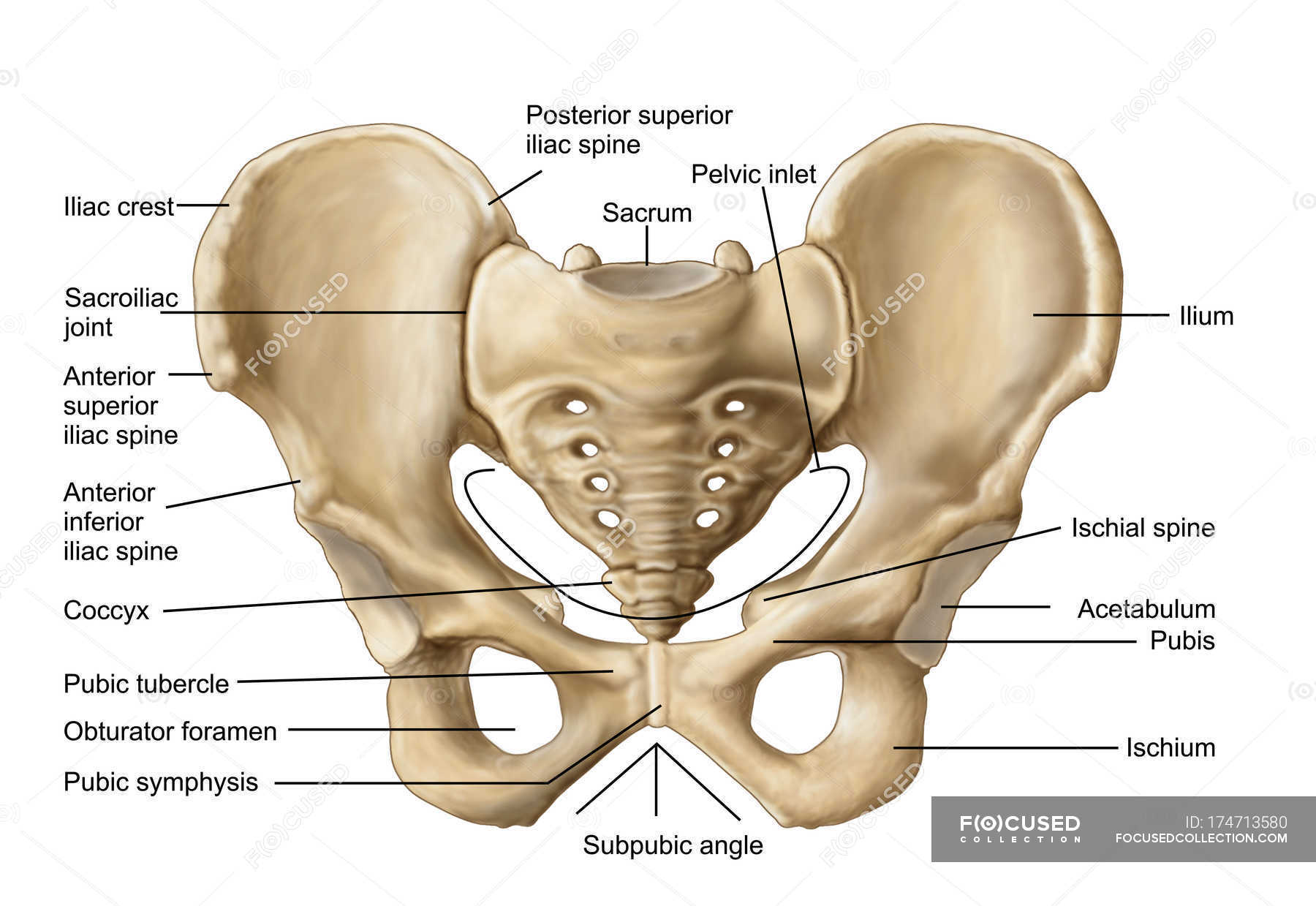
Anatomy Of Human Pelvic Bone With Labels Osteology Biology Stock Photo 174713580

Pelvic Girdle Posterior View With Labels Appendicular S Flickr
Pelvis Anatomy Recon Orthobullets

Human Skeleton System Pelvis With Labels Anatomy Stock Photo Download Image Now Istock
0 comments
Post a Comment